Background
Brain microglia (MG) are an important reservoir in PWH. We asked how frequently MG harbor proviruses, what fraction of these proviruses are intact, and where they integrate in the human genome. To address these questions, we obtained autopsy tissues from 3 PWH on suppressive ART enrolled in the “Last Gift” rapid autopsy program.
Methods
DNA was extracted from cell isolations of autopsy tissues, including brain (mixed tissue) MG (CD11b+ with 95.9% TMEM119+), spleen cells (bulk), brain T cells (CD3+), and spleen T cells (CD3+). DNA was diluted to a proviral endpoint in 384-well plates for multiple displacement amplification (MDA). MDA products were screened for potentially intact proviruses by PCR detection of LTR, PSI, and RRE, regions commonly deleted in defective proviruses. Integration site sequencing was performed on LTR+ wells to map host genome locations and assess clonality of infected cells.
Results
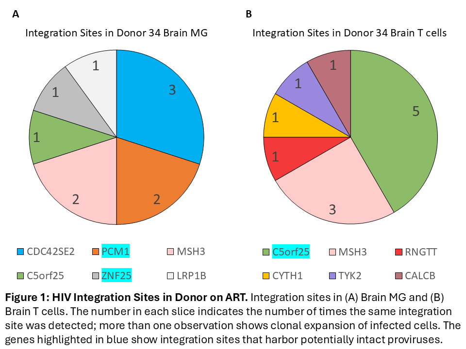
HIV proviruses were detected in both brain and spleen, the majority of which were defective. In one donor, 27% of MG-derived proviruses and 48% of spleen-derived proviruses were potentially intact (PSI+RRE+). We observed large clones through identical integration sites in both brain and spleen tissues, consistent with T cell origin (Fig. 1a, 1b). We also identified small clones observed in MG only, some of which are potentially intact, supporting a specific myeloid reservoir (Fig. 1a).
Conclusions
We identified infected MG harboring potentially intact proviruses and two highly expanded T-cell clones detected by integration site analysis that were present in brain MG, brain T cells, and spleen of PWH on ART. These findings suggest that infiltrating HIV-infected T cells contribute to brain proviral burden, potentially through attachment to or phagocytosis by MG. Defining the cellular reservoirs of HIV in brain is needed to target them for elimination.
Table or Figure