Background
People living with HIV (PLWH) on effective antiretroviral therapy (ART) continue to experience persistent immune activation and chronic inflammation in the absence of detectable viremia. This dysregulation contributes to increased risk of age-related comorbidities, which occur earlier and with higher frequency in PLWH compared to people without HIV (PWOH). It remains unclear, however, whether these complications are driven by pathways unique to HIV infection or represent an exacerbation of normal aging processes.
Methods
We used unbiased RNA-sequencing and high dimensional flow cytometry to examine CD4+ T cells from virus-suppressed “older” (≥55 years old) PLWH, “younger” (≤35 years old) PLWH, “older” PWOH, and “younger” PWOH. Sequencing reads were aligned using STAR. Telescope was used for single-locus transposable element (TE) mapping. Normalization and differential expression analysis was performed by DESeq2 and gene pathways were assessed using gene set enrichment analysis (GSEA). A 35-color panel was used to assess cell immunophenotypes by flow cytometry.
Results
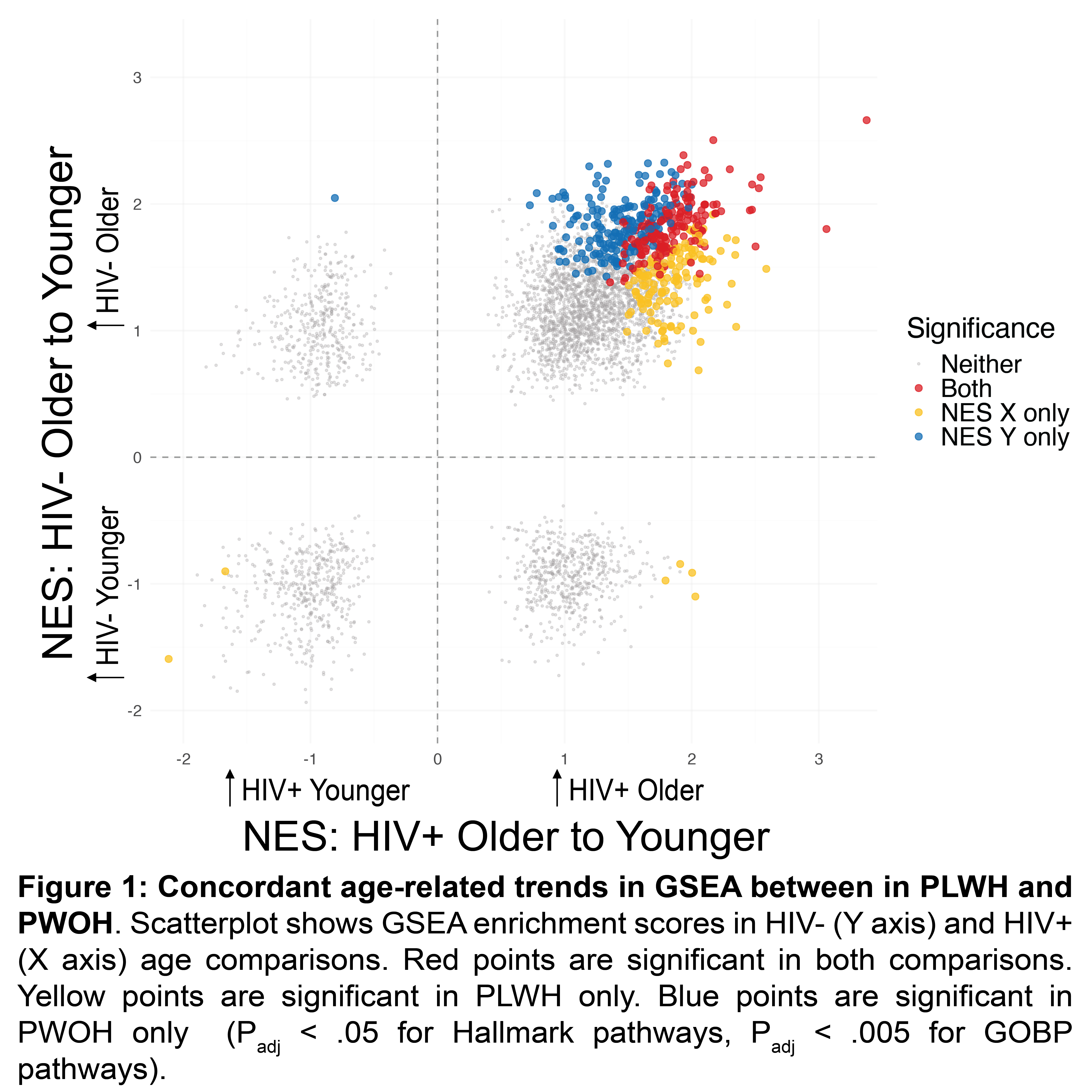
GSEA revealed broad overlap in pathways affected by aging, with 97% of pathways demonstrating concordant trends in comparisons between older versus younger PLWH and PWOH (Figure 1). Top pathways upregulated in older individuals regardless of HIV status included TNFα signaling (FDR = 6.21×10⁻³⁴), apoptosis (FDR = 1.30×10⁻¹¹), and T cell activation (FDR = 6.42×10⁻⁹). Fourteen percent of these age-related pathways were also significantly enriched in older PLWH compared with older PWOH, while no pathway was significantly upregulated in older PWOH compared to older PLWH. Among TE reads, a cluster of TEs on chromosome 2p12a were strongly downregulated with age in both PLWH and PWOH (log2FC = -2.22, p = 5.73 × 10⁻¹⁴). Expression of these TEs exhibited a strong negative correlation with several gene pathways related to regulated necrosis (ρ: -.844, p = 2.57×10⁻⁷), suggesting they may play a role in inflammatory cell death with age. Similar age-related changes were observed in flow cytometry, including increases in CD45RO+ memory T cells and TIGIT+ T cells, as well as a decrease in CD38+ T cells regardless of HIV status (all p < .005).
Conclusions
These results indicate that in CD4+ T cells, aging is marked by largely concordant transcriptional changes in PLWH and PWOH, supporting a model in which chronic HIV infection does not induce significant unique changes during aging but instead amplifies existing age-related programs.
Table or Figure