Background:
Among patients who begin DTG treatment an increase in mild to severe anxiety, insomnia, depression has been observed. Little is known about the mechanisms involved in these symptoms. Zebrafish emerged as an important model for drug screening and neurological research, due to its high similarity with mammals in neuro-anatomical and behavioural features. We explored the locomotor behaviour of zebrafish embryos exposed to DTG and the possibility of a folate rescue.
Methods:
Wild type and transgenic (tg:ngn1-EGFP) zebrafish embryos were exposed to 1 µM DTG with/without 5 µM folate from gastrula stage (4 hpf) up to 120 hpf. Locomotor activity was studied by analyzing wild-type larval swimming in a 2h trial period under dark/light cycling to analyze the embryos’ ability to adapt to environmental stimuli. To understand the relationship between altered behaviour and CNS development, we used tg:ngn1-EGFP embryos, expressing the green fluorescent protein under the promoter regulation of ngn1, a transcription factor playing an important role in the development of dopaminergic neurons.
Results:
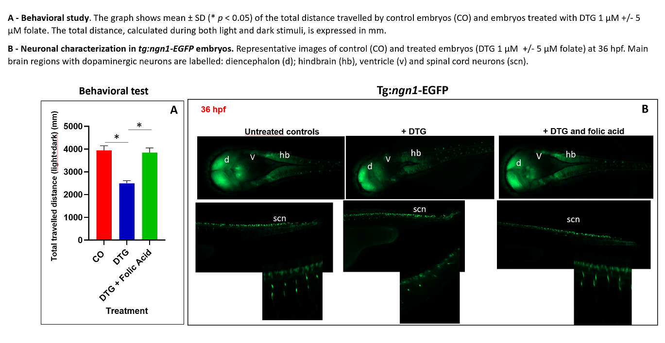
Embryos treated with DTG had significantly reduced locomotor activity (50% reduction). Folate addition completely restored movement frequency. ngn1-driven fluorescence was evident in control embryos in CNS regions corresponding to the diencephalon (d), hindbrain (hb), ventricle (v) and the trunk region within spinal cord neurons (scn). In treated embryos, ngn1 expression was decreased and impaired in those brain areas particularly enriched with dopaminergic neurons, like (d), (v) and (h), while a consistent number of (scn)s, peripheral projections of central dopaminergic neurons, were missing, as evidenced by the fluorescence pattern. The addition of folate restored the normal phenotype.
Conclusions:
The swimming behaviour of zebrafish embryos in light/dark stimuli is increasingly employed in studying neuroactive drugs. Locomotion in zebrafish is a complex behaviour produced by the activity of various neuronal pathways and its reduction upon the light/dark transition is interpreted as an anxiety state. Our results in the zebrafish embryo model show for the first time DTG effects on the dopaminergic pathways, characterized by a decrease in locomotor activity and an impaired expression of ngn1 in dopaminergic areas of the CNS and in the (scn)s of the trunk that can be rescued by folate co-administration.
Figure 1. Behavioral study and neuronal characterization in tg:ngn1-EGFP embryos.