Background:
Antiretroviral therapy (ART) initiation during acute HIV-1 infection limits HIV reservoir seeding and favors immune restoration, however there are concerns of remaining increased systemic inflammation, despite intervention with very early ART. In-depth analysis of lymph nodes during and after acute HIV-infection, may provide insight into the drivers of this inflammation.
Methods:
Samples were selected from a multi-centric Belgian cohort study (ACS), in which blood and inguinal lymph nodes (LNs) were collected during acute HIV infection (T0), upon reaching an undetectable plasma viral load on ART (UD) and 1 year later (UD+1). We performed multiplex immunoassay (Mesoscale Discovery) on plasma of 25 participants at T0 and UD. A total of 15 T0 and 8 UD+1 formalin-fixed paraffin embedded inguinal LNs were analyzed by multiplex immune fluorescence (IF) to determine inflammatory changes and activation. In addition, HIV DNAscope was used to quantify the frequency of HIV DNA+ (vDNA) cells. To maximize the sensitivity of the assay, coverage of subtype-specific probe sets was predicted based on near full-length sequences. In case < 20 ZZ pairs were predicted to bind, custom probe sets were used.
Results:
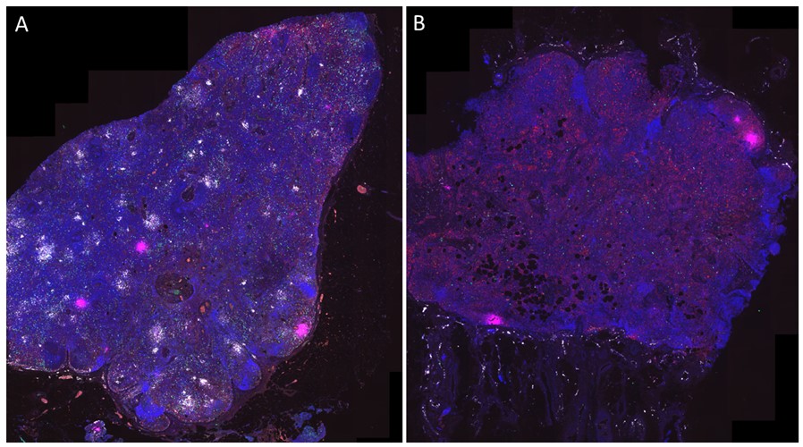
Plasma levels of CXCL10 and CXCL11 at UD were significantly higher in ACS-participants compared to healthy controls (p=0.018, p=0.015). CXCL10 was detected in a highly focal pattern within LNs and was positively correlated with pSTAT1 expression (r=0.614, p=0.001). Both showed a non-statistically significant downward trend between T0 and UD+1 (p=0.46, p=0.09) and foci of CXCL10 remained detectable in most LNs at UD+1. CXCL10-expressing cells consisted of 37.8% macrophages and 13.9% neutrophils at T0. Neutrophil counts within the LNs were elevated at T0, and were significantly reduced at UD+1 (p=0.031). Expression of CXCR3, the CXCL10 receptor, was significantly higher at T0 versus UD+1, in both T-cells, B-cells, and other cells (p=0.031), and was enriched in proliferating (Ki67+) cells. Initial quantification of vDNA showed a decrease between T0 ( median 482/106 cells) and UD+1 (median 75/106 cells), however no significance was reached (p=0.0625). No correlation could be found with expression levels of pSTAT1 or CXCL10.
Conclusions:
During acute HIV infection, lymph nodes are in a hyperinflammatory state. Foci of CXCL10 did not significantly decrease at UD+1, suggesting ongoing inflammation within the lymphoid tissues as a source for increased systemic CXCL10, despite suppressive ART.
Representative IF images on inguinal lymph nodes of an early treated participant (ACS005) for CXCL10 (magenta), pSTAT1 (white), neutrophil marker (MPO, green), pan-macrophage marker (Iba1, red) and nuclei (DAPI, blue) at the T0 timepoint (A) and UD+1 timepoint (B).