Previous studies have yielded conflicting results regarding the contribution of the gut to the overall CD4+ T-cell pool. SPECT imaging of whole-body CD4-pool in monkeys (by our group) using an anti-CD4 antibody (mAb) fragment (CD4R1-F(ab)’2) radiolabeled with Technetium-99m revealed low CD4-pool in the small and large intestines, corroborated by ex-vivo data. In contrast, PET imaging (by others) using the same anti-CD4R1 fragment radiolabeled with Copper-64 revealed substantial uptake in the gut of healthy animals and reduced uptake following SIV infection. The probe uptake in the clusters of lymph nodes and the spleen were similar between the two studies. Using a novel PET camera designed for monkeys (Mediso) with higher spatial resolution compared to clinical SPECT and PET cameras, we have repeated our experiments with the same anti-CD4R1 fragment radiolabeled with Zirconium-89 (89Zr).
The CD4R1-F(ab)'[sub]2[/sub] was radiolabeled with [sup]89[/sup]Zr using desferrioxamine with an isothiocyanate linker. Probe affinity was tested in MT4 cells. Healthy (n=6) or SIV-infected (n=3) rhesus macaques (CD4+T cells/µl: 90-2400) received intravenous injections of 58-127 MBq of [sup]89[/sup]Zr-CD4R1-F(ab)'[sub]2[/sub] (mAb-dose: ~100microg for five animals and ~1000 microg for the remaining four) and in-vivo imaged up to 6 days post-injection. PET-dynamic imaging coupled with kinetic modeling was performed to estimate binding potential (BP), the product of CD4 receptor molarity and ligand affinity.
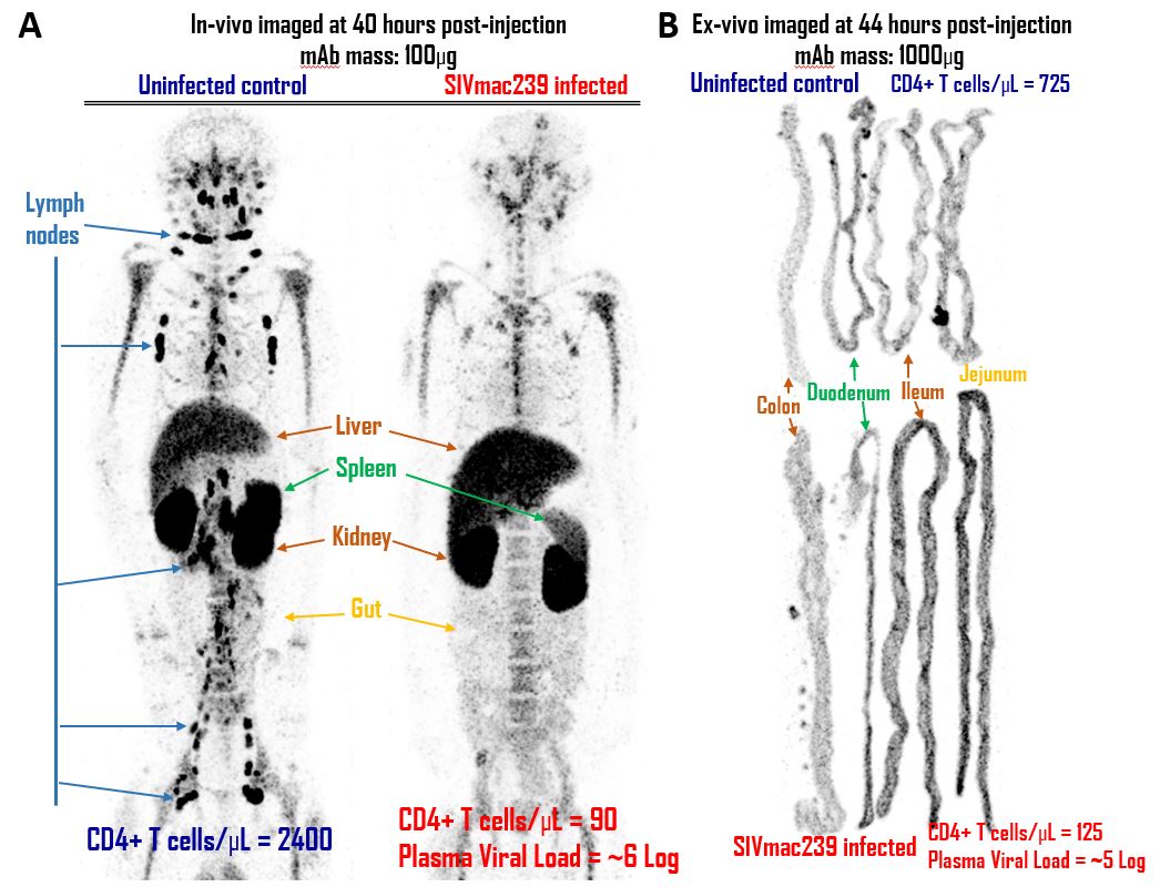
3D PET images of the CD4-pool revealed a clear delineation of lymph nodes and spleen, with dramatic differences of probe uptake in these organs between the healthy and the infected animals (Figure). Consistent with previous images obtained with 99mTc-CD4R1-F(ab)’2, regardless of the mAb dose, the new 89Zr-CD4R1-F(ab)’2 uptake in the gut was low and did not differ between healthy and SIV-infected animals. Ex-vivo studies of large and small intestines confirmed the in-vivo images. BP estimates showed the gut’s contribution to the total CD4 pool in humans to be lower than the spleen’s contribution alone.
The vast majority of specific binding to CD4+ tissue was localized to lymph nodes and spleen with minimal uptake in the gut. Gut uptake did not change in the setting of SIV infection. These data suggest that the prevalent notion of the gut as a major reservoir of the CD4 pool in the body needs to be revised.