Background
Young people with perinatal HIV (PHIV) have an increased risk of cardiac disease despite effective ART, particularly but not exclusively in those who have had delayed HIV diagnosis and antiretroviral therapy (ART) initiation. The aim of the study is to characterize the myocardial abnormalities in adolescents with PHIV who are established on ART using cardiac magnetic resonance (CMR) imaging and association with biomarkers of inflammation and cardiac stress.
Methods
We examined the myocardium in adolescents with PHIV aged 10-19 years taking ART compared to uninfected controls in Zimbabwe. Participants completed a 3-Tesla CMR assessment of cardiac function and myocardial fibrosis using late gadolinium enhancement (LGE). Circulating biomarkers of inflammation (CRP, TNF-α) fibrosis (Gal-3, TIMP-1) and cardiac stress (ST2, GDF-15) were assessed using multiplex bead assay (Luminex). Multivariate linear regression assessed how biomarker levels related to cardiac abnormalities. A p-value of 0.05 or less was considered significant.
Results
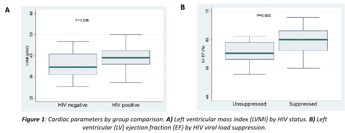
A total of 120 adolescents were enrolled (60 PHIV; 47% female and 60 HIV-uninfected; 42% female). Participants with HIV were older [median (IQR) 18 (16-19) vs 15 (13-17) years; p<0.001] compared to uninfected controls although both groups had a similar body surface area. In the HIV group, median age at HIV diagnosis was 5.5 (IQR) 4-8 years and 18 (82%) were virally suppressed (<19 copies/ml). 96 participants had CMR scans (49 PHIV and 47 uninfected controls). Both groups had similar myocardial function and no LGE. PHIV group had larger left ventricular (LV) mass index (LVMi) [39.4 (5.9) vs 35.9 (6.0) g/m2; p=0.006] . There was reduced LV ejection fraction [55.2% vs 59.9% ; p=0.005] and right ventricular ejection fraction [55.2% vs 59.2% ml/m2; p=0.026] in those who were not virally suppressed compared those suppressed. Plasma levels of TNF-α, TIMP1 and GDF-15 were higher in the PHIV group compared to uninfected control (p<0.001). CRP and ST2 were higher in those not virally suppressed versus unsuppressed. ST2 was significantly associated with higher LVMi (adjusted β, 1.368 ; p=0.040), LV end diastolic and systolic volume index (3.428 ; p=0.006 and 2.030 ; p=0.027).
Conclusions
CMR revealed similar myocardial function between PHIV and controls but mildly increased LVMi in the PHIV group. ST2 biomarker may suggest presence of subclinical myocardial stress. Longitudinal studies to understand the clinical implications of these myocardial alterations are required.
Table or Figure