Background
Factors that affect intact proviral DNA (IPD) decay in PWH receiving antiretroviral therapy (ART) are incompletely understood. We evaluated the effect of age, sex and markers of senescence on IPD decay, and examined individual decay slopes to assess whether there is heterogeneity in IPD changes.
Methods
Participants with long-term documented HIV suppression continuously followed for up to 25 years in the ACTG A5321 cohort had proviral DNA (intact, defective, total) measured longitudinally at 4-5 time points from year 5 to year 25 on ART. Senescence-associated secretory phenotype (SASP) biomarker levels were measured at the same time points. By-participant modeling of log-transformed IPD used censored-data linear regression.
Results
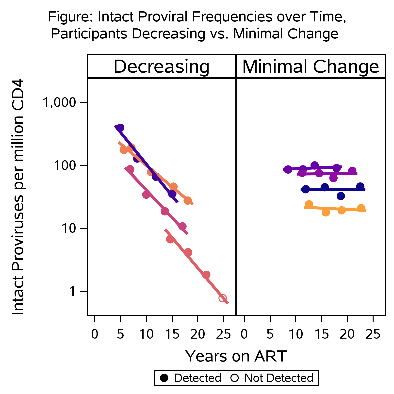
55 participants (27% female) were evaluated. Pre-ART median age, 43 years (range 22-60; IQR 32-48). In the overall population, IPD levels declined slowly after year 5 of ART (median half-life 8.6 yrs) whereas defective and total proviruses remained stable. Different patterns of IPD decay were observed. 32 participants (58%) had IPD slopes significantly different than zero and negative (indicating IPD decay; half-life 5.6 yrs). 4 participants with rapid decay (Figure) had IPD half-life of 3.3 yrs (8.3-fold reduction over 10 years). For the 23 participants (42%) who did not have significantly negative slopes, most had flat IPD slopes (minimal decay; examples in Figure). Three participants had apparent increase in IPD during long-term ART. At year 5 of ART, males had slightly higher median IPD than females (107 vs. 55 proviruses/million CD4 cells, p=0.13). Subsequent IPD decay was faster in males than females (half-life 7.5 vs. 35 yrs, p=0.028). 65% of males vs. 40% of females had significantly negative IPD slope. Levels at initial time point of two SASP biomarkers, MMP-2 and -9, did not correlate with IPD level at that time point or with subsequent IPD decay slope. MMP-2 and -9 levels also did not significantly correlate with age.
Conclusions
Intact proviral DNA decays slowly in most PWH on long-term ART. However, aggregate data can mask substantial differences in IPD decay across individuals followed longitudinally for 25 years. Whereas some PWH have minimal change in IPD after year 5 of ART, a subset continue to have significant IPD decay during long-term ART. Sex, but not age, is associated with IPD decay. Determining why some people have continued IPD decay is a high priority to inform future cure strategies.
Table or Figure